
Ralph Tufano, M.B.A., M.D.

https://www.hopkinsmedicine.org/profiles/results/directory/profile/0015647/ralph-tufano
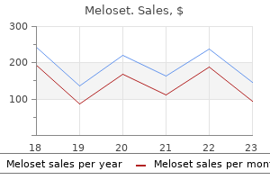
Meloset dosages:
Meloset packs:

The lymphatic vessels cross via nodes along the related colic artery to the central vessels on the colic artery root. Proximal to this the lymphatics drain along either the superior mesenteric artery (superior mesenteric nodes) or the inferior mesenteric artery (inferior mesenteric nodes) to the aorta/inferior vena cava (para-aortic nodes). The para-aortic lymphatics Types of specimen There are a wide variety of specimens, ranging from tiny biopsies through to elimination of the whole massive intestine. Most main colorectal resections may be carried out both open or laparoscopically, including robotic surgical procedure that has just lately been launched. Transverse colectomy Transverse colonic tumours could additionally be removed either by a wedge resection or by extending a proper or left hemicolectomy to embody the transverse colon. Left hemicolectomy/sigmoid colectomy the descending and/or sigmoid colon may be eliminated for most cancers or diverticular illness. Subtotal colectomy/total colectomy/ panproctocolectomy In cases of multiple tumours, hereditary polyposis or inflammatory bowel disease, surgeons might take away most of or the complete giant bowel with or with out the anal canal. Major rectal resections Anterior resection Anterior resection may be used to remove rectosigmoid or rectal tumours. The infra-peritoneal part of the rectum must be resected within the mesorectal fascial aircraft as described by Heald (total mesorectal excision) [11]. This can be extended to take away a half of the inner sphincter in very low tumours (intersphincteric dissection). The sigmoid colon could be anastomosed to the distal rectum or anal canal to restore intestinal continuity. Occasionally for anterior tumours where the margin is threatened, a cuff of vaginal wall or prostate may be taken with the specimen. Abdomino-perineal excision Abdomino-perineal excision of the rectum and anus is used for low rectal tumours close to the dentate line or those who contain the sphincters. The sphincters, with or with out the levator ani muscles, are eliminated en bloc with the mesorectum and anal canal: therefore the affected person requires a permanent colostomy. Occasionally, for giant advanced tumours involving multiple organs, a complete pelvic exenteration may be needed, the place all of the pelvic organs, and sometimes the Local colorectal excisions Polypectomy Colorectal polyps can be excised throughout colonoscopy by way of a diathermy snare handed around the stalk (if pedunculated) or sessile base. Preferably the specimen is eliminated entire; nevertheless, larger polyps or sessile lesions could also be removed piecemeal. Larger lesions ought to be serially sliced at 2- to 3-mm intervals and ideally embedded in their entirety. The polyp base must be inked if identifiable to affirm the completeness of excision. Endoscopic mucosal/submucosal resection these excisions could also be used for more sessile or flat adenomas, or for early cancers. The lesion is elevated by submucosal fluid injection and dissected off the submucosa with a small rim of regular mucosa. In the rectum, bigger lesions may be removed by transanal endoscopic mucosal resection. The specimen should be pinned out to forestall shrinkage and rolling of the perimeters throughout fixation. After inking the deep and lateral margins, the specimen should be serially sliced and embedded in separate cassettes. Cruciate sectioning could additionally be helpful to decide accurate distances to the lateral resection margin. Specimen handling and dissection Ideally, resection specimens should be received in the recent state instantly from the operating theatre, along with a request type confirming the affected person identity and full medical and specimen particulars. If an extended delay is anticipated or within the case of small biopsies, the specimen ought to be placed immediately in to an sufficient amount of formalin fixative, a minimal of 10 occasions larger than the tissue quantity. Examination of the contemporary specimen could be very helpful before fixation, significantly within the case of huge or complicated excisions. For non-neoplastic disease, the bowel could be opened earlier than fixation through the anterior peritoneal floor along the anterior taeniae coli. If the tumour is of a enough dimension it may possibly Normal massive gut 515 be everted via the proximal incision to take fresh materials for tissue banking if desired.
Syndromes
In addition, the authors counsel that some glutamate antagonists could also be helpful in decreasing the unwanted effects of ethambutol-a sensible suggestion that appears worthy of clinical investigation. The receptive fields in the left and right sides of space 17 mirror the contralateral visual world and representations of the higher and decrease areas of the visual area are separated below and above, respectively, the calcarine fissure. Cells in the posterior features of the calcarine fissure have receptive fields located in the central part of the retina. Cortical cells progressively deeper within the calcarine fissure have retinal receptive fields which would possibly be positioned increasingly peripherally within the retina. The central part of the fovea has tightly packed photoreceptors for decision of nice detailed images, and the cortical illustration of the central fovea is proportionately bigger than the peripheral retina to have the ability to accommodate a proportionately larger need for neural picture processing. The magnocelluar and parvocellular pathways project in a unique way to the histologically outlined layers of primary striate visible cortex and then to extrastriate visual areas. Cortical cells reply higher to traces of a specific orientation than to simple spots. The receptive fields of cortical cells are thought to characterize computational summaries of a quantity of simpler input signals. As the visible data proceeds from area V1 to extrastriate visible cortical areas, the illustration of the visible world reflected within the receptive fields of individual neurons becomes progressively more complex (Kaplan and Benardete, 2001). Ethambutol the dextro isomer of ethambutol is extensively used as an antimycobacterial drug for the remedy of tuberculosis. It is well-known that ethambutol produces dose-related alterations in the visible system, corresponding to blue�yellow and red�green dyschromatopsias, decreased distinction sensitivity, lowered visible acuity, and visual area loss. The earliest visual symptoms seem to be a lower in contrast sensitivity and colour vision, although impaired red�green colour imaginative and prescient is essentially the most frequently observed and reported criticism. However, the lack of contrast sensitivity could explain why some sufferers with normal visual acuity and shade notion nonetheless complain of visible disturbance. These visual system alterations can happen with a few weeks of doses equal to or larger than 20 mg/kg body weight; nonetheless, they normally turn into manifest after several months of therapy (Koliopoulos and Palimeris, 1972; Polak et al. The symptoms are primarily associated with certainly one of two types of retrobulbar optic neuritis (ie, optic neuropathy). The commonest kind, seen in virtually all instances, includes the central optic nerve fibers and typically results in a central or paracentral scotoma in the visual subject and is associated with impaired red�green color imaginative and prescient and decreased visual acuity, whereas the second form involves the peripheral optic nerve fibers and usually ends in a peripheral scotoma and visible area loss (Lessell, 1998; Bartlett and Jaanus, 2008). Pharmacological studies, using the in vivo and in vitro fashions, show that although Lead In addition to the well-documented retinal effects of lead (see above), lead exposure during maturity or perinatal growth produces structural, biochemical, and functional deficits within the visual cortex of humans, nonhuman primates, and rats (Fox et al. Quantitative morphometric studies in monkeys exposed to either high levels of lead from birth or infancy to six years of age revealed a decrease in visible cortex (areas V1 and V2), cell volume density, and a lower in the number of preliminary arborizations amongst pyramidal neurons (Reuhl et al. The former results may be due to an absolute decrease in total cell numbers, probably ensuing from lead-induced apoptosis as observed within the retina (Fox et al. This may account for the decreased density of cholinergic muscarinic receptors discovered in the visible cortex of grownup rats following reasonable stage developmental lead publicity (Costa and Fox, 1983). The morphometric outcomes on neuronal branching are harking back to earlier findings in the neocortex of rats following high level developmental lead exposure (Petit and LeBoutillier, 1979), and up to date findings in the somatosensory cortex of rats following low or moderate degree developmental lead publicity (Wilson et al. These alterations could partially contribute to the decreases in distinction sensitivity observed in lead-exposed rats and monkeys (Fox, 1984; Rice, 1998), the alterations in the amplitude and latency measures of the flash and pattern-reversal�evoked potentials in lead-exposed children, employees, monkeys, and rats (Fox et al. Methyl Mercury Methyl mercury became infamous in two episodes of mass poisoning (see Chap. In the 1950s, industrial discharges of mercury in to Minamata Bay in Japan became biomethylated to kind methyl mercury, which then accumulated within the food chain and reached poisonous concentrations within the fish and shellfish consumed within the surrounding communities. Hundreds of individuals had been poisoned, displaying a mix of sensory, motor, and cognitive deficits. Visual deficits are a prominent feature of methyl mercury intoxication in grownup humans, together with several other neurological manifestations corresponding to difficulties with sensation, gait, memory, and cognition. Methyl mercury poisoned people skilled a hanging and progressive constriction of the visual area (peripheral scotoma) as sufferers grew to become progressively much less in a place to see objects within the visible periphery (Iwata, 1977). The narrowing of the visual area provides impression of trying by way of an extended tunnel, therefore the term tunnel vision.
Churg�Strauss syndrome) Non-infectious Part of systemic involvement Inflammatory problems of the stomach a hundred and fifteen Table eleven. Endemic iron deficiency associated with Helicobacter pylori an infection among school-aged children in Alaska. Enhanced Fe ion-uptake activity in Helicobacter pylori strains isolated from sufferers with iron-deficiency anaemia. Association of Helicobacter pylori an infection and top of Mexican youngsters of low socioeconomic degree attending boarding schools. The organism possesses powerful urease activity, which is the idea for a quantity of biochemical exams for infection. There is considerable genomic diversity however pathogenicity relies upon to some extent on the production of varied potent proteins, especially vacuolating toxin (vac-A) and cytotoxin-associated protein (cag-A). Strains possessing these cytotoxins are associated with elevated inflammation and mucosal harm [62]. The higher prevalence among aged individuals reflects greater infection rates after they have been youngsters somewhat than an infection at later ages. Nevertheless, studies on water Histology the principal and most blatant histological function of H. In addition, variable numbers of neutrophils are current, characteristically found in and around the epithelium lining the base of gastric pits. In instances of severe inflammation, neutrophils are also current between and adjoining to the surface epithelial cells. Active inflammation in association with generalised diffuse gastritis is almost invariably related to the presence of H. Examination of semi-thin resin-embedded sections demonstrates that the epithelium shows cellular oedema, micropapillae, mucin loss and epithelial denudation. The elevated permeability of damaged surface epithelium could allow permeation of bacterial antigens and toxins and enhance underlying mucosal inflammatory response and harm [70]. Lymphoid follicles are a standard characteristic of helicobacter gastritis [71], particularly within the antral mucosa and in cases of extreme lively gastritis as compared with gentle or comparatively inactive cases [72,73]. Eradication of the organism results in a slow lower within the variety of lymphoid follicles they usually are inclined to lose their germinal centres and proof of lymphoid activation [74]. Marked histological lymphoid hyperplasia, sometimes leading to antral nodularity on endoscopy, is also a feature of H. Neutrophils disappear and epithelial cell damage heals within a matter of weeks [76�79]. The persistent inflammatory cell infiltrate, nonetheless, regresses extra slowly, especially in the antrum. Other workers have famous a slower disappearance of the mononuclear cell infiltrate stretching over a period of four years [78]. Eradication of the organism also results in a gradual decrease in the variety of lymphoid follicles [74]. Duodenal ulcer is often associated with antral predominant gastritis, little or no oxyntic gland (corpus and fundus) atrophy, and regular or increased acid secretion. Conversely, gastric ulcer and the intestinal sort of gastric cancer are sometimes associated with pangastritis, widespread oxyntic atrophy with varying levels of intestinal metaplasia, and hypo- or achlorhydria. Chronic inflammatory sequelae, similar to intestinal metaplasia, are for probably the most half (initially) confined to the antrum. A consistent discovering, related to marked duodenitis, occurring in sufferers with early H. The front progresses uniformly and so appears to advance sooner on the lesser curve. Thus, antral predominant gastritis might in some instances represent an earlier stage of atrophic pangastritis, these patterns representing two ends of the spectrum of H. Not solely that, but the organisms seem to migrate deeper in to the oxyntic glands, as opposed to their traditional superficial location. Histology continues to be thought of the gold standard for detecting the an infection both for untreated people and after remedy. However, histology is dear, actually in contrast with simple exams out there on the time of endoscopy, and a few have doubted whether the additional info offered by histological assessment, aside from confirming the presence of H pylori infection, is effective for the routine management of individual sufferers within the absence of an endoscopically detected lesion or lesions.

Two broad theories exist with regard to histogenesis: the primary proposes that they come up due to pancreatic maldevelopment [107]. This is supported by the frequent presence, throughout the tumour, of misplaced pancreatic tissue and the high incidence of immunoreactivity for pancreatic polypeptide and somatostatin. The second principle suggests a neoplastic lesion [108], with a complex triphasic development pattern. Rare reports of native lymph node deposits composed of the endocrine cell component, both alone [111] or with sparse spindle cell or ganglion cell components [110], help this. An similar tumour occurs within the cauda equina area [112], the origin of which is difficult to clarify on the basis of pancreatic maldevelopment. Poorly differentiated endocrine carcinoma/small cell carcinoma Fewer than 30 circumstances of this uncommon and highly aggressive periampullary tumour have been recorded in the literature, most presenting with obstructive jaundice, stomach pain and weight loss, and adopted by a quickly fatal course [114,115]. The tumours are small (20�30 mm), ulcerated or protuberant lesions which microscopically reveal sheets or nests of small, mitotically active cells with round or oval hyperchromatic nuclei, scanty cytoplasm and foci of necrosis. Using haematoxylin and eosin (H&E) staining, they resemble both small cell carcinomas or massive cell endocrine carcinomas. Malignant lymphoma must be excluded by immunocytochemistry and metastatic tumours by medical examination and radiology. Localised tumours could also be resectable and metastatic tumours may reply to chemotherapy [72,117]. Goblet cell carcinoid A few cases of this rare tumour have been reported throughout the duodenum, either at the ampulla of Vater [118,119] or inside the duodenal bulb [120]. Morphologically, these were small tumours that appeared equivalent histologically to their extra frequent counterparts within the appendix (see Chapter 30), being composed of small infiltrative nests of cytologically bland goblet cells, signet-ring cells and endocrine cells. By analogy with appendiceal goblet cell carcinoids, their prognosis should be intermediate between these of properly differentiated endocrine tumours and adenocarcinomas of the duodenum. The neoplasm has an aggressive behaviour and dying usually results from widespread metastases [125]. One examine showed loss of expression of p14, p15 and p16 in the 9p21 gene cluster [139]. Small tumours are sometimes discovered incidentally post mortem or at laparotomy whereas larger neoplasms current with belly ache, intestinal obstruction or ischaemia from the local results of the tumour. In addition, these tumours frequently lead to a dense desmoplastic response of the mesentery, usually at a distance from the first tumour; this will likely cause subacute or frank obstruction. Multiple tumours are related to a younger age of onset, an elevated danger of carcinoid syndrome and a poorer prognosis [141]. This final discovering appears to be consistent with the present view that a quantity of tumours might symbolize metastases as opposed to independent major neoplasms. A current examine [142] showed an equivalent X-chromosomal inactivation pattern in multiple tumours, implying a standard neoplastic clone. Palliative surgical procedure is individualised on a case-by-case basis: resection of an asymptomatic major tumour, even within the presence of liver metastases, is recommended to stop small bowel occlusion or local issues at a later stage [143]. The main lesions in the bowel wall range from barely palpable foci of thickening to nodules measuring as a lot as 35 mms; rarely do the primary tumours exceed this measurement. Clinically, patients may current with intestinal obstruction, volvulus or ischaemia. It would appear to be a result of the impact on blood vessels by the tumour(s) [147] (see Chapter 23). Elastic�van Gieson staining reveals adventitial elastic sclerosis of each mesenteric arteries and veins, with luminal venous thrombosis. Ghosts of necrotic tumour cell islands (yellow) are seen among the many perivascular fibrous tissue. The tumour cells are uniform, with little pleomorphism, nuclear hyperchromasia or mitotic activity. A new proposed grading scheme using mitotic count and Ki-67 index provides the potential for increased prognostic stratification (Table 24. Erosion of the serosal surface might lead to solitary peritoneal deposits [148] or to a diffuse peritoneal carcinomatosis [149]. Involved lymph nodes may become massive, measuring as a lot as 60 mm in diameter, and are frequently considerably bigger than the first tumour within the bowel wall.
Genetic and environmental factors in age-related nuclear cataracts in monozygotic twins. A neuropsychological study of children with elevated dentine lead degree: assessment of the effect of lead in different socioeconomic teams. Differences in color imaginative and prescient impairment caused by digoxin, digitoxin, or pengitoxin. Bcl-xL overexpression blocks baxmediated mitochondrial contact web site formation and apoptosis in rod photoreceptors of lead-exposed mice. Lead and calcium produce photoreceptor cell apoptosis by opening the mitochondrial permeability transition pore. Epidemic optic and peripheral neuropathy in Cuba: a unique geopolitical public health downside. Electrophysiological evaluation of complicated mind techniques: sensory evoked potentials and their mills. The influence of vision on computerized neurobehavioral check scores: a proposal for bettering take a look at protocols. Studies on the effect of 4-methylpyrazole on retinal activity within the methanol poisoned monkey by recording the electroretinogram. Chronic optica-neuropathy due to environmental publicity of organophosphate pesticides (Saku disease): clinical and experimental research. Neuro-ophthalmological findings and a follow-up research within the Agano space, Niigata Pref. Visual short-term results of Viagra: double-blind examine in healthy younger topics. Optic atrophy with visible subject defect in workers occupationally uncovered to lead for 30 years. Retinal degeneration and different eye problems in wives of farmer pesticide applicators enrolled in the Agricultural Health Study. Pattern visual evoked cortical potentials in patients with poisonous optic neuropathy brought on by toluene abuse. Persistent lower of the dopamine-synthesizing enzyme tyrosine hydroxylase within the rhesus monkey retina after chronic lead exposure. On acquired color imaginative and prescient disturbances during treatment with ethambutol and indomethacin. Investigation of discrepancy between darkish adaptation and electroretinographic findings in advanced stages. Diminished regulation of mesolimbic dopaminergic exercise in rat after persistent inorganic lead exposure. Vision disorders and phosphodiesterase 5 inhibitors: a evaluation of the evidence to date. A rat model manifesting methanol-induced visible dysfunction appropriate for each acute and long-term publicity studies. Toxicological analysis of preservative-containing and preservative-free topical prostaglandin analogues on a three-dimensional-reconstituted corneal epithelium system. Persistent will increase in scotopic b-wave amplitudes after lead publicity in monkeys. Alteration of the visual evoked potential and the electroretinogram in lead-treated monkeys. Selective degeneration of the parvocellular-projecting retinal ganglion cells in a New World monkey, Saimiri sciureus. Electrophysiological and electroretinographic proof for phototreceptor dysfunction as a poisonous effect of digoxin. Hypoxia-induced manufacturing of 12-hydroxyeicosanoids within the corneal epithelium: involvement of a cytochrome P-4504B1 isoform. An overview on the toxic morphological changes in the retinal pigment epitheliu after systemic compound administration. Oxygen consumption in rat outer and inside retina: light- and pharmacologically induced inhibition. Substrate-dependent effects of calcium on rat retinal mitochondria respiration: physiological and toxicological studies. Chromal focus of acquired chromatic discrimination loss and slovent exposure among printshop staff.
Commiphora erythraea (Myrrh). Meloset.
Source: http://www.rxlist.com/script/main/art.asp?articlekey=96567

It is said to the likelihood of malignant transformation [11�13], and also to the danger of synchronous and metachronous adenomas [14]. Small lesions 5�10 mm diameter have been proven to have superior features in 10% and carcinoma in nearly 1%, so that they have a low but definite malignancy threat. The majority of adenomas eliminated, over 80�90%, are tubular adenomas <10 mm [16,17]. Adenomas Epidemiology the prevalence of typical adenomas varies throughout the world. The Paris endoscopic classification of superficial neoplastic lesions: esophagus, stomach, and colon: November 30 to December 1, 2002. In a follow-up examine of barium enema-detected polyps >10 mm discovered earlier than the routine use of colonoscopy, 37% of the polyps elevated in size and 10% of sufferers developed carcinoma at the web site of the polyp over a mean period of 108 months [18]. In that study, actuarial evaluation advised an 8% carcinoma risk at 10 years and 25% at 20 years. In addition, 5% of sufferers developed a colorectal adenocarcinoma remote from the unique polyp. Macroscopically, adenomas have historically been described as pedunculated or sessile. In these lesions, foci of irregular ulceration should raise the risk of carcinoma arising within the adenoma. Standardised definitions are missing [21] but the elevation is usually lower than the height of closed biopsy forceps (2. A color difference compared with the surrounding mucosa may be the solely macroscopic change. These adenomas can unfold laterally, becoming giant and carpet like; others probably evolve in to polypoid adenomas [23]. When central melancholy is mixed with a raised edge, the malignant threat is especially excessive [24,25]. Truly flat and depressed adenomas are troublesome to detect, typically displaying only slight congestion macroscopically. The mouths of crypts from tubular adenomas can be recognised by chromoendoscopy as rounded openings which would possibly be larger than the traditional adjacent crypts. Macroscopic modifications that may be recognised embody haemorrhage, torsionassociated congestion, ulceration or diathermy change from the polypectomy. Prolapsing pedunculated adenomas could present pseudo-invasion with mucin or mucosal displacement in to the stalk, inflicting thickening on this space as properly as mucinous cysts and extravasates on the cut face. Microscopic appearances Adenomas come up as single crypts lined by dysplastic epithelium (monocryptal adenomas), in all probability following mutation or epigenetic gene silencing in colonic stem cells situated on the crypt bases [27,28]. With continued proliferation of dysplastic epithelium, adenomas are believed to grow by both crypt fission with vertical branching and development of the irregular epithelium in to adjacent crypts [28,29]. Aberrant crypt foci can be detected in situ by excessive magnification chromoscopic colonoscopy and are characterised by an altered form of the luminal opening, thickened epithelium and larger than normal crypts [30]. Increased numbers of those foci have been described in sufferers who even have adenomas or carcinoma [31]. Microscopically, two key features are used to outline and classify typical adenomas � the architecture and the diploma of dysplasia (intraepithelial neoplasia). The architectural sample assesses the proportion of tubular elements, characterised by epithelial glands surrounded by lamina propria, and villous components by which the epithelial lining incorporates the lamina propria. Thus, the tubular and villous components in an adenoma may be likened to the structure of crypts and villi in the small gut. Other authors have suggested a cut-off of 25% villous elements [16] however the distinction is somewhat semantic because the evaluation is made by estimation. Polyps and tumour-like lesions of the large intestine 651 at all times be simple because of issues in figuring out whether constructions are villi or open tubules [1], in addition to the need to estimate the degree of villosity. It has been noted that the reproducibility of this classification primarily based on the proportion of villous elements is imperfect [33,34] and at best solely average [35,36], but an attempt is inspired due to the significance of classification in figuring out the long run neoplastic danger and follow-up screening intervals [37].
Gastric inflammatory fibroid polyp handled with Helicobacter pylori eradication remedy. Concomitant presence of inflammatory fibroid polyp and carcinoma or adenoma in the stomach. Lipid islands in human gastric mucosa: morphological and immunohistochemical findings. Adenomatous transformation in hamartomatous polyps cases of two patients with Peutz-Jeghers syndrome. Cowden syndrome: report of two circumstances and evaluate of medical presentation and management of a uncommon colorectal polyposis. Generalized gastrointestinal polyposis: an uncommon syndrome of polyposis, pigmentation, alopecia and onychotrophia. Cronkhite� Canada syndrome with colon cancer, portal thrombosis, excessive titer of antinuclear antibodies, and membranous glomerulonephritis. Cronkhite�Canada syndrome associated with superior rectal cancer treated by a subtotal colectomy: report of a case. Gastric xanthomatosis associated with gastric intestinal metaplasia in a dyspeptic affected person. Fine-needle aspiration cytology findings from a case of pancreatic heterotopia at the gastroesophageal junction. Mucinous cyst exhibiting extreme dysplasia in gastric heterotopic pancreas related to gastrointestinal stromal tumour. Adenocarcinoma arising in gastric heterotopic pancreas: clinicopathological and immunohistochemical study with genetic evaluation of a case. Cytomegalovirus and Helicobacter pylori co-infection in a baby with M�n�trier illness. M�n�trier illness manifested by polyposis within the gastric antrum and coexisting with gastritis cystica profunda. Spectrum of hypertrophic gastropathy: large rugal folds, polyposis, and carcinoma of the stomach � case report and evaluation of the literature. Reversible proteinlosing hypertrophic gastropathy: causal relationship with Helicobacter pylori infection, or easy coincidence Immunolocalization of remodeling growth factor-alpha in normal and diseased human gastric mucosa. M�n�trier disease and gastrointestinal stromal tumors: hyperproliferative problems of the stomach. It is characterised by mobile atypia reflective of irregular differentiation and disorganised glandular structure. Determination of the proper prognosis and grade of dysplasia is crucial because it predicts the risk of both malignant transformation and metachronous gastric cancer [1]. In the early Nineteen Eighties, dysplasia was defined as unequivocally neoplastic epithelium [2] and graded in a two-tier system: low grade and high grade dysplasia. However, diagnostic standards and grading schemes have advanced in another way worldwide and there are disagreements between western and Japanese pathologists [3,4]. Non-invasive excessive grade neoplasia, together with three different sorts of lesions: excessive grade adenoma/dysplasia, non-invasive carcinoma (carcinoma in situ) and suspicion of invasion 5. Invasive neoplasia, together with intramucosal carcinoma in addition to carcinoma invasive in to the submucosa or beyond. The up to date model, offered in 2003, takes in to account enchancment in endoscopic methods and their administration implications. Consequently, non-invasive, high grade, pre-malignant lesions, without invasion of the lamina propria, and invasive adenocarcinomas confined to the lamina propria, have been placed in the single diagnostic class four (Table 13. The distinction between regenerative atypia and low grade intra-epithelial neoplasia/dysplasia could be difficult and is reflected in the vital inter-observer variation reported in the prognosis of dysplasia. In such instances, the dilemma may be solved by slicing to deeper ranges, obtaining extra biopsies or considering all attainable aetiologies. Foveolar hyperplasia might show irregular and tortuous tubular structures with epithelial mucus depletion, a high nucleus:cytoplasm ratio and loss of mobile polarity. Enlarged, hyperchromatic nuclei related to outstanding mitoses, if present, are often positioned close to the proliferative zone within the mucus�neck area. The glands might seem closely packed and lined by cells with large, hyperchromatic, basally situated nuclei.

The position of glial cells and apoptosis of enteric neurons in the neuropathology of intractable sluggish transit constipation. Acquired myopathic pseudo-obstruction could also be because of an autoimmune enteric leiomyositis. Gastrointestinal manifestations of progressive systemic scleroderma based mostly on a review of 364 instances. Progressive systemic sclerosis of the gastrointestinal tract and hereditary hole visceral myopathy: two distinguishable problems of intestinal easy muscle. Smooth muscle denervation in people is associated with inclusion physique formation in the gastrointestinal tract. Alimentarytract ganglioneuromatosis: a serious element of the syndrome of a quantity of endocrine neoplasia, Type 2b. Intestinal pseudoobstruction because the presenting manifestation of small cell carcinoma of the lung: a paraneoplastic neuropathy of the gastrointestinal tract. Procidentia of the rectum studied with cineradiography: a contribution to the discussion of causative mechanisms. Intussusception of the rectum � inner procidentia: remedy and leads to 90 sufferers. Impalement and anorectal injuries in childhood: a retrospective examine of 12 cases. Gall-stone obstruction of the sigmoid colon with specific reference to aetiology. Complications and therapy of migrated biliary endoprostheses: a evaluation of the literature. Mucosal prolapse syndrome � a unifying concept for solitary ulcer syndrome and related disorders. Submucosal mucous cysts at a colostomy website: relationship to colitis cystica profunda and report of a case. Pathology of the rectal wall in solitary rectal ulcer syndrome and complete rectal prolapse. Diseased mucosa can often be visualised and biopsied utilizing rigid procto-sigmoidoscopy, the versatile sigmoidoscope or the colonoscope. Bacterial culture of faeces is easy, although not all the time delicate, and numerous parasites could be demonstrated in faeces or mucus on direct microscopy. Bacterial infections that mimic persistent inflammatory bowel disease clinically are those that invade the mucosa. The commonest of those are those brought on by Clostridium difficile and Salmonella, Shigella and Cam pylobacter spp. In all inflammatory situations of the colorectum, the artwork is within the interpretation of the lesions. Inflammation as a outcome of viruses Although acute gastrointestinal an infection is a serious reason for morbidity all through the world and viruses play a leading half in its aetiology, viral infection of the colorectum rarely comes to the eye of the practising histopathologist. In truth, in most acute viral infections of the intestines, the small gut is the primary seat of infection. For a fuller description of viral an infection within the intestines, the fascinated reader is referred to Chapter 20. In this chapter, solely these viral infections with cytopathic effects demonstrable in histopathological sections of colorectal mucosal biopsies are considered. Among transplant recipients its incidence is highest in bone marrow transplant recipients occurring in about 5�20% of patients, kids being extra susceptible [2]. For the histopathologist, probably an important associations are inflicting lymphoid hyperplasia within the terminal ileum and subsequent intussusception in infants [3]. The adenovirus inclusions are intranuclear and eosinophilic with perinuclear halo, much like herpes virus, and are readily demonstrable in colorectal epithelium by immunohistochemical techniques. Cytomegalovirus the function of this herpes group virus as a major colonic pathogen is disputed.
The innocent indigenous small intestinal flora could become supplanted by irregular bacterial colonisation in numerous circumstances, including stagnant loops and fistulae. Although not essentially invasive, or inflicting structural changes visible in haematoxylin and eosin (H&E)-stained sections, such infections injury the glycocalyx, producing a partial lack of disaccharidase enzyme activity, leading to maldigestion. The surgical resections and anastomoses that native disease could require can themselves produce related effects. Local illness is commonly related to local mucosal ulceration, typically with some distortion of the adjoining mucosa. Malnutrition because of unsatisfactory food plan Protein�calorie malnutrition in children Protein�calorie malnutrition (usually generally recognized as kwashiorkor) results from extreme dietary protein deficiency and is present in Asia and in many African tribes, significantly in east Africa and South Africa. It impacts youngsters and adults but, in humans and experimental animals, epithelial adjustments are extra severe within the period of progress than in grownup life or old age [9]. In severe protein� calorie malnutrition, jejunal and ileal mucosal peak, floor space and the entire quantity of the lamina propria are all reduced. The mucosal flattening could be extreme in youngsters, typically mimicking gluten-induced enteropathy. The appearances counsel poor alternative of the conventional enterocyte loss as a outcome of failure in crypt cell proliferation, rather than an over-rapid lack of enterocytes [12]. Surgical resections the typical size of the small bowel in a stay adult is about 2800 mm. Earlier studies advised that a few third of the upper small bowel might be resected with impunity and half with reasonable safety [14] and that survival is feasible after a resection of 90% [15]. Children appear to tolerate extra intensive resections better than adults, notably with regard to their long-term results [16]. A single case of folate deficiency is reported [13] with flattening which improved and finally disappeared on dietary folate. Food intolerance Up to 20% of the populace of western countries understand an antagonistic reaction to a number of meals, and objectively 6% of infants and young youngsters and three. A genetic predisposition may exist and the pathogenesis involves a T-cell-mediated immune response, for which coeliac disease is the prototypical example, an IgE-mediated (often eosinophil-rich) allergic reaction, similar to that induced by shellfish or peanuts or a variable combination of those two. Cell-mediated immune issues account for most food intolerance-related malabsorption. A defect in the digestive enzyme methods, particularly those related to enterocytes, which can be inherited or acquired, the latter by publicity to an infection, drugs or different poisonous substances. The situation in these patients may be legitimately described as gluten-induced (or sensitive) enteropathy. The essential illness manifestation is the development of a characteristic enteropathy. However, coeliac illness might affect other parts of the gastrointestinal system and indeed different organ systems. It is evident from population-based serotesting that the illness prevalence is much larger than beforehand appreciated, with seropositivity approximating 1% in folks of European ancestry [21,22], although disease seems to be uncommon in African and east Asian populations. Clinically diagnosed coeliac illness is far much less common than the 1% prevalence suggested by serology, leading to hypothesis Aetiology and pathogenesis [22,24,25] the illness involves the complicated interaction of an environmental set off (gluten protein derived from the Triticeae tribe of grains), host elements (genetic predisposition) and environmental co-factors (enteric infection). Gluten is a generic time period encompassing grain proteins rich in prolamine and glutamine amino acids that confer pathogenicity for coeliac disease. These proteins embrace gliadins and glutenins from wheat, hordeins from barley and secalins from rye. Prolamine confers resistance to acid and enzymatic digestion, allowing a high focus of minimally altered gluten protein to enter the proximal small gut. Gluten features entry to the lamina propria by way of transcellular, paracellular and retrotranscytosis routes, with permeability to gluten peptides enhanced by factors such as enteric an infection with adenovirus or rotavirus and upregulation of zonulin, a decent junction regulator [22]. In the lamina propria, gluten peptides trigger both innate and adaptive immune responses. These induce enterocyte cell dying by Fas�Fas ligand interplay or perforin granzyme process. Matrix metalloproteinase action additionally contributes and the mix of these components leads in time to the villous flattening typical of coeliac illness. Chemoattractant cytokines are answerable for mast cell, eosinophil and neutrophil infiltration. Gluten-induced enteropathy Three parts have been necessary for complete certainty in prognosis; an irregular pattern on biopsy, enchancment in both scientific signs and histological mucosal morphology after gluten withdrawal and the reappearance of enteropathy after its re-introduction [21,23]. Histological modifications are assessed by means of endoscopic biopsies taken from the duodenum and proximal jejunum.

Within the physique mucosa, capillaries lengthen at right angles to the surface and kind a community beneath the surface epithelial layer surrounding the gastric pits, which is drained by sparse amassing venules. In the antral mucosa, there are two distinct capillary beds: a basal capillary system branching out from quick arterioles and a superficial capillary network originating from long arterioles ascending in to the mucosa [10]. Their absence from the superficial mucosa correlates with the low prevalence of lymph nodal metastases in intramucosal gastric most cancers [63,64]. The muscularis mucosae consists of the inner round layer and outer longitudinal layer, and accommodates some elastic fibres. In the antrum, vertical skinny fascicles extend up in to the mucosa and separate pyloric glands. Submucosa the submucosa is a thick layer of unfastened connective tissue beneath the muscularis mucosae, with comparatively thick arteries, veins and lymphatics forming main plexuses, as well as nerves and the submucosal nerve plexus of Meissner. The submucosa is wealthy in elastic fibres and likewise incorporates fibroblasts, adipocytes, easy muscle cells and mast cells. The internal round coat surrounds the whole abdomen and is steady with that of the oesophagus. The outer longitudinal coat runs from the oesophagus to the duodenum and is continuous with their longitudinal fibres. At the pylorus, the internal round muscle thickens and forms distinct proximal and distal loops which unite in a torus along the lesser curvature, forming the pyloric sphincter. Subserosa and serosa the subserosa is a thin layer of free areolar tissue containing blood vessels, lymphatics and nerve fibres. It is enclosed by the serosa, composed of a sheet of flattened mesothelial cells steady with the serosal lining of the peritoneal cavity. When infected, the mesothelial cells easily show reactive swelling and nuclear atypia, which should not be misinterpreted as malignant cells, particularly in peritoneal cytology [13]. Neural networks of the gastric wall and interstitial cells of Cajal the abdomen is richly innervated by each extrinsic and intrinsic nervous systems [66]. The extrinsic system contains the parasympathetic and sympathetic autonomic nerve supplies mentioned above, which include parasympathetic and sympathetic motor neurons and accompanying sensory neurons. The intrinsic system types correct nerve plexuses (submucosal plexuses of Meissner and myenteric plexuses of Auerbach). Integration of those two components allows the abdomen to respond to both local and exterior stimuli by regulating motility, native blood move, secretion, endocrine functions and mucosal defence mechanisms. In the wall, wealthy networks of cholinergic nerves are observed and an analogous network is current for gastric glands in the mucosa, whereas adrenergic neurons are scantier [67]. Other types of neurons are also current within the gastric mucosa, such as peptidergic (peptide-containing) and nitrergic (nitric oxide synthetase-containing) neurons [66]. In the muscle coat, interstitial cells of Cajal are interposed between easy muscle cells and neurons. They are pacemaker cells that regulate autonomous motility of the sleek muscle cells, additionally taking half in a task in mediating neurotransmission [68,69]. Double-contrast upper gastrointestinal radiography: a sample method for ailments of the stomach. Comparative examine on persistent gastritis with dissecting microscopic findings of stained abdomen and histological findings: application to endoscopic prognosis. Microgastroscopic findings of mucosal microvascular structure as visualized by magnifying endoscopy. Gastric microvascular structure as visualized by magnifying endoscopy: physique and antral mucosa without pathologic change demonstrate two totally different patterns of microvascular architecture. Clinical which means of pepsinogen take a look at and Helicobacter pylori serology in the well being check-up population in Korea. Histopathology of the gastrooesophageal junction: a examine on 36 operation specimens. The mucin profiles of regular gastric mucosa, intestinal metaplasia and its variants and gastric carcinoma. Gastric and intestinal combined and solely intestinal forms of intestinal metaplasia in the human stomach. A research of 3406 gastrectomy specimens from dwellers of the Atlantic and the Pacific basins.
References
Desarrolló: Grupo Guadalupe S.R.L.