
Cindy L. O�Bryant, PharmD, BCOP
http://www.ucdenver.edu/academics/colleges/pharmacy/Departments/ClinicalPharmacy/DOCPFaculty/H-P/Pages/OBryantCindyPharmD.aspx
Hyzaar dosages: 50 mg, 12.5 mg
Hyzaar packs: 30 pills, 60 pills, 90 pills, 120 pills, 180 pills, 270 pills, 360 pills
The left lateral section of the liver is elevated by dividing the left triangular ligament. Therefore the lesser omentum and left gastric artery ought to be separated from the lesser curvature on the degree of the stomach. The splenic artery is split to the left of the midline, far from the celiac axis, after which dissected towards the aorta, so that it can be rotated to the proper for exposure of the superior mesenteric artery, which lies deep to it. After a Kocher maneuver, the duodenum and pancreatic head are elevated and retracted caudally to expose the superior mesenteric artery, which is then dissected all the way down to the aorta. Care is taken not to transect an accessory or replaced proper hepatic artery by avoiding dissection on the right facet of the superior mesenteric artery. An aortotomy is carried out between the superior mesenteric and proper renal arteries and prolonged to present the arterial patch. The infrahepatic inferior vena cava is then transected simply above the renal veins. Meticulous in situ dissection in search of a right department can significantly improve extraction time. The kidneys could be stored en bloc for machine perfusion or separated and despatched directly to the recipient facilities. The method requires consensual, preextubation (premortem) femoral vessel cannulation. After declaration of death, chilly preservation answer is instantly infused via the femoral artery cannula, and the femoral vein cannula is opened to gravity to decompress the venous system. Thereafter, median sternotomy and midline abdominal incisions are made, and the intraabdominal organs are topically ice cooled after which eliminated en bloc or separately. To stop resumption of circulation to the heart and brain, an occlusion balloon catheter is introduced in the contralateral femoral artery and inflated within the descending aorta. In 2010 Guarrera et al78 reported the primary potential examine of human liver transplantation using organs preserved with ex vivo hypothermic machine perfusion, demonstrating its References Premortem Cannulation Technique, With or Without Extracorporeal Membrane Oxygenation the premortem cannulation technique, described by Groth and colleagues in Stockholm, Sweden74 and later by the group in Madison, Wisconsin,eight decreases the rush 17-19, 23,25, 26, fifty nine. In 2012 Boehnert et al79 showed that normothermic ex vivo perfusion reduces liver and bile duct harm of pig livers retrieved after cardiac demise. Maintain a low threshold for performing a donor liver biopsy to exclude centrilobular necrosis and different predictors of poor graft quality. Use T tubes liberally to facilitate (1) analysis of potential early postliver transplantation cholestasis and (2) surveillance for ischemic cholangiopathy. Pitfalls embody inadequate planning and poor communication with the donor coordinator and operating room nurses. Conflicts of curiosity embody involvement in decisions about affected person prognosis, withdrawal of support, or willpower of dying. After procurement, cautious assessment of information on the flow sheet is crucial for appraising the ischemic harm. Department of Health and Human Services, Health Resources and Services Administration, Healthcare Systems Bureau, Division of Transplantation, 2012. Utilization, outcomes, and retransplantation of liver allografts from donation after cardiac demise: implications for further growth of the deceased-donor pool. Donation after cardiac demise as a strategy to increase deceased donor liver availability. Events in procurement as threat elements for ischemic cholangiopathy in liver transplantation using donation after cardiac dying donors. The impression of ischemic cholangiopathy in liver transplantation using donors after cardiac dying: the untold story. Renal transplantations carried out using non-heart-beating organ donors: going again to the longer term. Outcomes of kidneys from donors after cardiac dying: implications for allocation and preservation. A comprehensive risk quantification rating for deceased donor kidneys: the kidney donor risk index.
This provides the benefit of excellent hemostasis and the flexibility to probe the biliary tree to delineate the anatomy rigorously earlier than division of the hilar structures. In contrast, ex situ splitting requires no further sources or surgical expertise at the retrieval, and the liver may be cut up whereas making ready the recipients for implantation. The majority of energetic splitting facilities are presently utilizing ex situ splitting techniques. The long-term outcomes of cut up liver transplantation are actually similar to these of whole grafts in both grownup and pediatric recipients Table 52-2)22,23. A thorough understanding of the surgical anatomy of the liver and notably that pertaining to the caudate lobe is crucial before enterprise split liver transplantation. Donor Selection the necessity for predictable liver operate and thus a goodquality liver was recognized early in the growth of break up liver transplantation. Donors lower than 40 years of age had been arbitrarily defined as appropriate for splitting to provide a left lateral phase graft for a child. Mildly fatty livers (less than 20%) may be suitable if cold ischemic times are saved as quick as attainable. Donors with mildly irregular liver operate (aspartate aminotransferase stage less than three times normal or if greater with a falling trajectory), intensive care keep of higher than 5 days, and significant vasopressor help can be used with caution. Short warm and cold ischemia occasions are important components to be thought-about if early dysfunction or ischemic cholangiopathy are to be prevented. The quantity of liver allotted to the child is calculated by the donor-recipient ratio of 10:1 (for example, a liver from a 70-kg donor could produce a left lateral section to be utilized in a 7-kg youngster approximately). Ex Situ Split Ex situ splitting is carried out after the donor liver has been retrieved and perfused with preservation resolution. It ought to be split within the transplant center on a back table ready for this purpose. Crushed sterile ice ought to be replaced regularly within the container to maintain the specified temperature. Regular drainage of water and substitute of ice helps to maintain the temperature at 4° C. A systematic evaluation of the next structures must be carried out: · Hepatic veins: the left hepatic vein ought to join the vena cava separately from the middle hepatic vein. As little dissection as attainable must be performed between the bile duct and the hepatic artery to protect the arterial provide to the bile duct. The allocation of vascular structures is dependent upon the liver anatomy and the transplant recipient. The portal vein is dissected to the bifurcation, and the tributaries to the caudate lobe are ligated and divided. If there are multiple arteries present, then allocation should be performed in such a way as to reduce the vascular reconstruction required. Once the vessels and bile duct have been allotted and divided, the dissection of the liver parenchyma starts at a point 1 cm to the proper of the falciform ligament. The parenchyma is crushed gently with a hemostatic clamp revealing vascular constructions, that are tied or clipped individually after which divided. The left lateral segment graft is then flushed with preservation resolution by way of the artery and portal vein to test leakage at the cut surface. Further cut-surface sutures are often required to achieve passable hemostasis. The vessels/structures for this graft are the left hepatic vein, hepatic artery (left hepatic artery or common hepatic artery), portal vein (left or main trunk), and left hepatic duct. Careful probing of the bile duct is useful in gaining understanding of the anatomical variations of the biliary tree. Resection of the caudate lobe and oversewing of the portal plate reduces the potential for bile leaks from unrecognized caudate bile ducts. Again, cautious and delicate dissection of the porta hepatis must be performed to determine anatomical variations distal to the origin of the gastroduodenal artery. Identification of left or right accent hepatic arteries from the left gastric or superior mesenteric artery, respectively, is pretty simple during dissection, and their presence will determine the allocation of the vascular trunk to every graft. The allocation of the main vascular constructions in the absence of variant arterial anatomy (main trunk of the portal vein or the common hepatic artery) will depend upon the traits of the recipients and expertise of the transplant team.
Diseases
The infrahepatic vena cava is sewn in precisely the identical manner as used for the suprahepatic vena cava, usually with 4-0 polypropylene, and a 1. Excessive length of vena cava is the main reason for folding and kinking, which may cause formidable postoperative problems that require extraordinarily difficult reconstructions at a later stage. A laparotomy pad is placed against the right hemidiaphragm to prevent kinking of the portal vein anastomosis. If dimension have to be adjusted to both the donor or the recipient portal vein, a fish-mouth reconstruction is really helpful. It is our preference to release the portal vein clamp slowly with a watch on the electrocardiogram to monitor the procedure. We normally open the portal vein in phases after any T-wave phase elevations have reversed. It usually takes three or four partial decompressions earlier than the vein is totally open. In such instances, arterial reconstruction and reperfusion of the liver are performed before the portal conduit is sewn in. Otherwise, the anastomosis line might rupture fully and cause an immediate catastrophe. Sometimes, giant collateral veins may be discovered within the subhepatic area, both alongside the minor curvature of the stomach or as a collateral to the liver, that can be used for the donor portal vein anastomosis instead of a mesenteric venous conduit. If necessary, we proceed to venovenous bypass or carry out an end-to-side portocaval shunt. The portal vein is rotated slightly counterclockwise, which facilitates its approximation to the vena cava. With the inferior liver floor utterly uncovered, the dissection is sustained within the aircraft between the cava and the liver. Once the liver is removed, all clips and ligatures are secured by 3-0 or 4-0 silk sutures. The left and middle veins are clamped and divided to enable the liver to be removed from the wound. When dividing the proper, middle, and left hepatic veins, we divide them as far into the liver as practical. Depending on the anatomy, we join the hepatic veins into a standard cloaca and extend the cava incision distally three to four cm. The donor cava is minimize down longitudinally in the again 3 to 4 cm from the proximal cava to create two corresponding orifices. It is easily done by placing the proper lobe down into the hepatic fossa and lifting the left lobe as much as expose the 2 cavas. A stitch at each finish of the anastomosis (cranial, caudal) makes it easy to outline the suture line for completion. At this point the portal vein reconstruction is performed in regular style (see earlier). For reperfusion we usually start by opening the portal vein and flushing air and intravascular high-potassium fluid out of the liver by way of the open finish of the distal portion of the donor cava. The distal end, controlled by two keep sutures, is clamped when the flush seems sufficient. At this time the recipient cava clamp is opened to set up regular venous return. The distal finish of the donor cava is oversewn with working 4-0 monofilament suture. The arterial anastomosis is sewn with 6-0 or 7-0 polypropylene, and a one-half-diameter development factor is tied in place to provide full growth of the arterial anastomosis without any constriction. The purpose for this sequence in selected circumstances is to prevent a malrotation of the changed right hepatic artery that can occur if the reconstruction is performed on the back desk. In some sufferers, despite an ideal technical result, inflow is inadequate because of compression of the celiac axis by the arcuate ligament. This complication is demonstrated by marked respiratory variation and could be documented by flow measurement. In this situation the celiac artery have to be dissected proximal to the aorta and the arcuate ligament minimize. Quite incessantly within the case of twin blood provide to the liver from an accessory proper hepatic artery and a hepatic artery proper, the accent right hepatic artery stemming from the superior mesenteric artery is dominant. In such instances the donor celiac artery is anastomosed to the recipient accessory proper hepatic artery.
Exogenous anions will also improve the focus of unmeasured anions (salicylate toxicity, ethylene glycol, and methanol ingestion). Hyperchloremic acidosis and renal tubular acidosis (bicarbonate loss) could have a traditional anion gap. Measurement of the plasma lactic acid focus and calculation of the anion gap from sodium, chloride, and bicarbonate permits differentiation of dilutional acidosis from acidosis due to tissue hypoperfusion. Excess administration of sodium bicarbonate may be an iatrogenic reason for metabolic alkalosis. Compensation for Acid�Base Disturbances Respiratory acidosis is compensated for inside 6 to 12 hours by elevated renal secretion of hydrogen ions, with a resulting increase within the plasma bicarbonate focus. After a quantity of days, the pH shall be regular despite persistence of an increased Paco2. Metabolic acidosis stimulates alveolar air flow, which causes rapid elimination of carbon dioxide from the physique and decreases the hydrogen ion focus towards regular. This respiratory compensation for metabolic acidosis, however, is simply partial because pH remains somewhat beneath normal. As with metabolic acidosis, the respiratory compensation for metabolic alkalosis is just partial. Renal compensation for metabolic alkalosis is elevated by reabsorption of hydrogen ions. Chapter 26 � Acid�Base Disorders 615 this metabolic compensation is limited by the availability of sodium, potassium, and chloride ions. During prolonged vomiting, there could additionally be extreme lack of chloride ions together with sodium and potassium. When this happens, the kidneys preferentially conserve sodium and potassium ions and the urine turns into paradoxically acidic. Effects of Temperature on Acid�Base Status Temperature modifications have a number of effects on blood and tissue pH and Pco2. Therefore, for a given carbon dioxide content material, the partial pressure will lower because the temperature falls. The blood pH is additional elevated because the dissociation of water into protons and hydroxyl ions decreases with cooling, thus reducing hydrogen ion concentration. In addition, proton buffering by hemoglobin a-imidazole teams is enhanced by hypothermia. These adjustments are in all probability insignificant inside the physiologic temperature range however are important when deciphering blood-gas and acid�base knowledge during induced cooling throughout cardiopulmonary bypass. Two alternate blood-gas administration methods, "a-stat" and "pH-stat" are utilized throughout hypothermia in the working room (Table 26-6). During hypothermic cardiopulmonary bypass, this technique normally includes the addition of carbon dioxide by way of the oxygenator. A purported advantage of this technique is that cerebral blood move shall be increased because carbon dioxide is a po- tent cerebral vasodilator. Temperature correction of blood gasoline samples is required to interpret the values obtained from a hypothermic patient but measured at 37�C. The pH-stat technique is used more typically in surgery for pediatric congenital coronary heart disease, especially throughout cooling and deep hypothermic circulatory arrest. Cerebral harm secondary to world hypoperfusion is thought to be a larger menace than supply of microemboli on this patient population. Hypothermia, hypocarbia (via the Bohr effect), and alkalosis, all shift the oxyhemoglobin dissociation curve to the left and impair tissue oxygen supply. The addition of carbon dioxide during pH-stat administration will counter these effects and facilitate oxygen unloading from hemoglobin. Homeotherms and hibernating animals hypoventilate in order to preserve blood pH at 7. However, the mind and coronary heart of those animals make use of a-stat methods to maintain pHi at a-stat values and to hold up near normal perform. Poikilotherms use the a-stat strategy and allow their blood pH to increase and Pco2 to lower with cooling to find a way to preserve mobile and enzyme perform over wide temperature ranges.
They may be a results of sluggish circulate inside a small however patent vessel, high-grade stenosis, or technical failure. A, Arteriogram reveals high-grade stenosis on the hepatic artery anastomosis (arrow). Real-time ultrasonographic assessment of the liver reveals a heterogenous appearance within the liver parenchyma with multiple discrete areas of low echogenicity, findings according to hepatic ischemia or infarction (+). B, Celiac axis arteriogram demonstrates occlusion of the hepatic artery proximally (arrow). Intrahepatic flow may be detectable from the arterial collaterals in the porta hepatis. It is necessary to avoid this diagnostic error as a end result of delay in prognosis could end in lack of the allograft. When detected late, symptoms often relate to ischemic biliary injury and manifest as recurrent cholangitis, intrahepatic infarction and abscess, bile duct stenosis, or bile leak. Presentation as fulminant hepatic necrosis is uncommon when ultrasonography is used for postoperative monitoring. Early analysis may permit emergency surgical thrombectomy or hepatic artery reconstruction, or each, which could stop or postpone the need for retransplantation, or serve as a bridge until a donor turns into out there. Because thrombolysis with tissue plasminogen activator can entail a danger for bleeding, we suggest that the affected person stay within the angiography suite while the infusion is happening in order that extravasation could be detected. Intrahepatic false aneurysms might happen following a percutaneous liver biopsy or transhepatic biliary drainage. Some false aneurysms are asymptomatic and detectable only on imaging; others might manifest as gastrointestinal bleeding, hemobilia, or hemoperitoneum. On Doppler ultrasonography they appear as spherical fluid collections displaying inside flow. The scientific manifestations of portal vein thrombosis are variable and embody graft failure, ascites, varices, and gastrointestinal bleeding. Significant stenosis can normally be detected by duplex and colour Doppler ultrasonography. Portal vein stenosis is recognized on Doppler ultrasonography by the presence of normal proximal flow, a high-velocity jet on the anastomosis, and distal turbulence. Air within the portal vein has sometimes been detected on Doppler ultrasonography as highly echogenic, nonshadowing particles transferring inside the portal vein. Although air was reported in one case in association with a gangrenous colon,eighty five it may be a transient finding without other serious complication in the first 2 weeks after transplantation. Celiac angiogram reveals a extreme stenosis of the hepatic artery with quite a few collaterals within the porta hepatis. Confusion of these collaterals with the main hepatic artery could result in erroneous interpretation of the ultrasound research and end result within the false-positive interpretation that the hepatic artery is patent. A, Flush aortogram reveals the stump of an occluded donor iliac artery graft from the infrarenal aorta to the liver transplant (arrow). A gentle irregularity consistent with residual thrombus is seen within the hepatic artery. The portal vein is entered through a transhepatic puncture; after entry to the portal system is achieved, pressures are measured throughout the stenosis and balloon angioplasty is carried out. Success was obtained in two sufferers, each of whom had been asymptomatic at 1 12 months follow-up. We routinely perform portal vein angioplasty in patients, normally youngsters who develop portal vein stenosis after transplant. Olcott et al88 described their expertise with balloon angioplasty in four sufferers with portal vein occlusion. Three of the four treated sufferers died of a quantity of problems unrelated to the angioplasty. The researchers additionally described the location of a steel stent in a affected person with portal vein occlusion. This affected person died of a brain abscess 1 month later, and the stent was patent at post-mortem.
Progression to thrombosis may lead to liver infarction, abscess, or biloma formation. The early clinical indicators of vascular occlusion are often nonspecific, and differentiation from other causes of graft dysfunction, corresponding to acute rejection, an infection, and prolonged chilly or heat preservation ischemic injury, may be difficult. A, Selective celiac artery injection reveals an apparent gentle stenosis on the anastomosis of the donor and recipient hepatic arteries (arrow). B, A digital subtraction arteriogram with oblique angulation of the picture intensifier exhibits the stenosis to be severe (arrow). A smaller photopenic region is recognized in the left lobe of the liver (short arrow). In suspected hepatic artery obstruction, arteriography must be performed to confirm the ultrasonographic or cross-sectional imaging findings. Once a stenosis is detected angiographically, the therapy indicated depends on the scientific signs and liver function. Stenoses occurring within the quick postoperative period are usually caused by technical problems and generally require operative revision. Recurrent stenosis was reported by Raby et al65 in hepatic artery anastomoses at 4 and 6 months in two of three patients; both stenoses responded to another balloon dilation. There is limited experience with endovascular hepatic artery stent placement, but in selected instances they are often positioned. Flint et al72 reported the correct diagnosis of hepatic artery thrombosis with Doppler ultrasonography in 34 of 37 cases (92%); the diagnoses were confirmed by angiography or surgery. In three cases the outcomes have been false-positive, and hepatic arteries that appeared patent with Doppler ultrasonography proved on angiography to be occluded. Hall et al73 noticed false-negative Doppler ultrasonography studies in 4 of 13 circumstances (31%). In a big sequence of more than 2200 transplantations, Zajko et al59 observed 10 cases of stenosis and two cases of thrombosis. The "piggyback anastomosis" (with preservation of the recipient vena cava and cavocaval anastomosis) could lead to venous obstruction. Delayed caval stenosis may be as a outcome of fibrosis, a persistent thrombus, or neointimal hyperplasia. Pressure measurements must be obtained in the inferior vena cava and across the hepatic veins. A, Superior mesenteric artery incompletely subtracted; V, superior mesenteric vein. The infrahepatic portion of the inferior vena cava is attenuated; nevertheless, contrast materials drained freely into the right atrium, no gradient was detected, and the ultrasonography study was normal. In uncomplicated cases the dimensions of the ducts tends to remain steady over time; nonetheless, a mild improve in the measurement of the ducts may occur with out scientific proof of obstruction. Diffuse biliary tree adjustments are often seen in the absence of obstruction or leakage. These are nonspecific findings that are seen with rejection, hepatitis, preservation harm, and infarction. T tubes placed within the recipient bile duct at the time of liver transplantation are removed 2 to four months after surgical procedure. If the cholangiographic findings reveal no abnormality, the T tube is eliminated over a guidewire, and a quick lived drain is placed within the tract to avoid leakage of bile into the peritoneum. Simple removal of the T tube utilizing commonplace strategies can outcome in bile leakage. At our establishment, sufferers developed bilomas and critical bile peritonitis following easy removal of the T tube. Therefore a small-caliber, multiplesidehole, straight catheter is positioned within the T-tube tract. This straight catheter is withdrawn progressively over the course of 24 to forty eight hours.
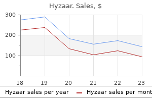
Kreteks (Clove). Hyzaar.
Source: http://www.rxlist.com/script/main/art.asp?articlekey=96275
Metoclopramide within the prevention of postoperative nausea and vomiting: a q uantitative systematic evaluate of randomized, placebo-controlled research. Interventions for stopping nausea and vomiting in girls undergoing regional anaesthesia for caesarean part. Comparison of intermittent versus steady infusion metoclopramide in command of acute nausea induced by cisplatin chemotherapy. Comparison of intermittent ondansetron versus continuous infusion metoclopramide used with standard mixture antiemetics in command of acute nausea induced by cisplatin chemotherapy. Postoperative neurologic dysfunction associated with preoperative administration of metoclopramide. Akathisia and anesthesia: refusal of surgery after the administration of metoclopramide. Extrapyramidal side effects after metoclopramide administration in a post-anesthesia care unit- a case report. Dose-dependent effect of metoclopramide on cholinesterases and suxamethonium metabolism. The use of H1 and H2 histamine antagonists with morphine anesthesia: a double-blind study. The association between cognition and histamine-2 receptor antagonists in African Americans. The hemodynamic effects of intravenous cimetidine in intensive care unit patients: a d oubleblind, potential research. Intragastric acidity, micro organism, nitrite, and N-nitroso compounds before, during, and after cimetidine remedy. Does pretreatment with cimetidine and ranitidine affect the disposition of bupivacaine Effect of famotidine and lansoprazole on serum phosphorus levels in hemodialysis patients on calcium carbonate remedy. Short-term treatment with proton pump inhibitors, H2-receptor antagonists and prokinetics for gastro-oesophageal reflux disease-like symptoms and endoscopy negative reflux disease. A comparability of lansoprazole, omeprazole, and ranitidine for lowering preoperative gastric secretion in adult sufferers present process elective surgical procedure. Influence of metoclopramide on plasma cholinesterase and period of motion of mivacurium. The antibiotic azithromycin is a motilin receptor agonist in human stomach: comparison with erythromycin. Intravenous erythromycin dramatically accelerates gastric emptying in gastroparesis diabeticorum and normals and abolishes the emptying discrimination between solids and liquids. Motilin agonist erythromycin will increase human lower esophageal sphincter strain by stimulation of cholinergic nerves. The use of erythromycin as a gastrointestinal prokinetic agent in grownup critical care: advantages versus risks. Parenteral vitamin is defi ed as delivery of vitamins instantly into the venous circulation (peripheral vein or central vein). Although the advantages of parenteral diet within the perioperative interval are controversial, postoperative enteral feeding has been proven to decrease complication charges in malnourished sufferers although mortality rates are unchanged. For instance, power necessities might double and protein necessities might triple in severely burned patients. Minimally careworn patients require about 25 to 30 cal/ kg and 1 g/kg of protein daily to stay in nitrogen and vitality equilibrium. Moderately to severely careworn patients should be resuscitated first after which started on a hypocaloric regimen (20 cal/kg) till the stress response abates. Lipid calories from infusions of propofol may be important and ought to be included when calculating caloric intake. Th ee a long time ago, it was thought that the principle aim of vitamin support within the hospitalized patient was to satisfy power requirements and to make the patient anabolic. Current objectives embrace meeting and attenuating the metabolic response to emphasize and, as properly as, attenuating cellular harm and modulating the immune response to damage.
Evaluating fibrinogen ranges represents a critical facet of all transfusion algorithms, especially for patients with huge transfusion and life-threatening hemorrhage. Transfusion algorithms are a cr itical and relatively new facet of perioperative administration; they attempt to offer sufficient issue and hemostatic substitute, although the perfect ratio of assorted blood components and factor concentrates are nonetheless being decided. Murray Liver the liver lies in the right upper quadrant of the stomach cavity and is hooked up to the diaphragm. It is the biggest organ within the physique, weighing approximately 1,500 g a nd representing 2% of body weight. Hepatocytes characterize approximately 80% o f the cytoplasmic mass within the liver. The capacity of hemopoietic stem cells to distinguish into hepatocytes introduces the risk of treating inherited disorders of metabolism (reflecting absent to altered enzymes because of a single or a number of genetic defect) sooner or later. Endothelial cells that line the hepatic lobules contain massive pores, permitting simple diffusion of certain substances, together with plasma proteins, into extravascular spaces of the liver that join with terminal lymphatics. The excessive permeability of the lining of endothelial cells permits giant portions of lymph to kind, which comprise protein concentrations which would possibly be solely slightly less than the protein concentration of plasma. Indeed, approximately one-third to one-half of all of the lymph is shaped in the liver. Total hepatic blood circulate is roughly 1,450 mL p er minute or approximately 29% o f the cardiac output. Of this quantity, the portal vein supplies 75% of the whole circulate however only 50% to 55% of the hepatic oxygen provide because this blood is partially deoxygenated in the preportal organs and tissues (gastrointestinal tract, spleen, pancreas). The hepatic artery offers solely 25% of complete hepatic blood move but provides 45% to 50% of the hepatic oxygen necessities. Hepatic artery blood fl w maintains nutrition of connective tissues and partitions of bile ducts. For this purpose, loss of hepatic artery blood flow may be fatal because of ensuing necrosis of vital liver buildings. An increase in hepatic oxygen necessities is met by an increase in oxygen extraction quite than a further increase in the already high hepatic blood move. Anatomy the liver is split into four lobes consisting of 50,000 to a hundred,000 particular person hepatic lobules. Blood flows past hepatocytes through sinusoids from branches of the portal vein and hepatic artery to a central vein. There is usually just one layer of hepatocytes between sinusoids so the whole area of contact with plasma is great. Central veins be part of to kind hepatic veins, which drain into the inferior vena cava. Each hepatocyte can be situated adjacent to bile canaliculi, which coalesce to form the widespread hepatic duct. This duct and the cystic duct from the gallbladder join to kind the common bile duct, which enters the duodenum at a site surrounded by the sphincter of Oddi. Hepatic lobules are lined by macrophages (derived from circulating monocytes) generally identified as Kupffer cells, which phagocytize 99% or more of bacteria in the portal venous blood. This is crucial because the portal venous Control of Hepatic Blood Flow Portal vein blood flow is controlled primarily by the arterioles in the preportal splanchnic organs. Sympathetic nervous system innervation is from T3 to T11 and is mediated through a-adrenergic receptors. This innervation is principally liable for resistance and compliance of hepatic venules. Changes in hepatic venous compliance play a vital role in total regulation of cardiac output and the reservoir perform of the liver (see the section "Reservoir Function"). Fibrotic constriction characteristic of hepatic cirrhosis (most usually due to persistent alcohol abuse and hepatitis C) can improve resistance to portal vein blood flow, as evidenced by portal venous pressures of 20 t o 30 m m Hg (portal hypertension). Ascites results when elevated portal venous pressures trigger transudation of protein-rich fluid via the outer surface of the liver capsule and gastrointestinal tract into the abdominal cavity. Hepatic artery blood circulate is influenced by arteriolar tone that reflects native and intrinsic mechanisms (autoregulation). For example, a d ecrease in portal vein blood flow is accompanied by a rise in hepatic artery blood move by as much as 100 percent. Chapter 32 � Gastrointestinal Physiology 671 subsequent hepatic arterial vasodilation and washout of the vasodilating material. Halothane decreases hepatic oxygen supply greater than isoflurane, enflurane, desflurane, or sevoflurane when administered in equal potent doses.
Hepatic venous congestion in living donor liver transplantation: preoperative quantitative prediction and follow-up utilizing computed tomography. Living donor liver transplantation in adults: Vascular variants necessary in surgical planning for donors and recipients. Variants of the bile ducts: medical software within the potential donor of living-related hepatic transplantation. The left bile duct (white arrow), proper bile duct, and common bile duct (gray arrow) are properly visualized. Imaging of hepatic arterial anatomy for depicting vascular variations in living related liver transplant donor candidates with multidetector computed tomography: comparison with conventional angiography. The recipient was a Japanese boy who received a left lateral segment from his mother. Unfortunately, persistent rejection developed a yr later, and the affected person underwent retransplantation with a liver from a deceased donor. As the next step, an revolutionary technique of splitting an adult graft was developed and has turn into increasingly refined. Pediatric recipients were most well-liked for 2 causes: dad and mom could be most simply chosen as potential donors with the fewest moral problems, and donor security may be more easily preserved by leaving the larger proper lobe of the donor intact after harvesting the left aspect of the liver. The left lobe is normally enough for the metabolic calls for of a pediatric recipient. Split-liver transplantation from deceased donors, if sufficiently out there, can cut back the variety of pediatric patients ready for organ donation without decreasing the persistently inadequate donor pool for grownup sufferers. This stability is affected by the availability of deceased donors, so this situation ought to be completely different in every country. In Japan the connection between the donor and the recipient is regulated by the principle of the Japanese Society of Transplantation that the donor and recipient should be relations Table 48-1). However, in some cases, social stress may oblige the dad and mom to turn out to be donors. Grandparents could additionally be additionally included among the many donor candidates, and in such circumstances further risk to an aged donor should be included within the knowledgeable consent dialogue after meticulous evaluation of the well being of the donor candidate. When the donor is a extra distant relative, such as an uncle or aunt, voluntary willingness ought to be rigorously ascertained. In common, donor candidates ought to be interviewed independently within the absence of the recipient and different relations. Complications and long-term end result of living liver donors: a survey of 1508 in five Asian facilities. Adhesion of the gastric wall to the reduce surface of the liver additionally occurs more frequently after left-sided hepatectomy than after proper lobectomy. Donor candidates must be knowledgeable in advance about all the attainable donor dangers. Family members or physicians and coordinators should clarify the remedy before the process. They could also be distressed to live with the sacrifice being made by their mother or father or relative. They may expertise appreciable anxiety concerning the operation and the lengthy run if not utterly knowledgeable. For instance, instances with malignant disease similar to hepatoblastoma, which is giant and invasive, could additionally be considered to have low priority or be excluded from listing in deceased donor transplantation. B, Computed tomography reveals the hairpin curve of the gastric outlet on the adhesion site (arrow). Suitable donors can be selected on the premise of two views: (1) donors with normal liver function and normal anatomy with sufficient dimension of the liver and (2) donors without any systemic diseases, abnormalities, and high risk of postoperative risks. If multiple candidates can be found, conventional research could additionally be carried out at the preliminary screening for each if requested. Such studies embody a blood depend; coagulation profile; blood chemistry studies for hepatic and renal operate; serological exams for hepatitis C and B virus, syphilis, and human immunodeficiency virus; electrocardiography; chest radiography; and ultrasonography of the liver. After preliminary examination the outcomes are offered as a software for selecting the ultimate candidate. The age of the donor ought to ideally be between 20 and 60 years, though we include 60 to 70 years if no youthful donor is out there. Younger candidates must be evaluated for their competence in making a voluntary decision.
Renal illness disrupts this regulation, and ectopic tissue calcification in addition to hyperphosphatemia may outcome. Profound skeletal muscle weakness sufficient to contribute to hypoventilation could also be attributable to hypophosphatemia, in addition to central nervous system dysfunction and peripheral neuropathy. Causes of hypophosphatemia include alcohol abuse; extended parenteral diet; medicines similar to acetazolamide, catecholamines, and theophylline; paracetamol overdose; large burns; restoration from hypothermia; hemodialysis; salicylate poisoning; and gram-negative bacteremia. Approximately 80% of the iron in plasma enters the bone marrow to be included into new erythrocytes. In addition to bone marrow, iron is incorporated into reticuloendothelial cells of the liver and spleen. Iron can also be an integral part of many enzymes needed for energy switch. Hemoglobin synthesis is the principal determinant of the plasma iron turnover rate. When blood loss happens, hemoglobin concentration is maintained by mobilization of tissue iron shops. Indeed, hemoglobin concentrations turn into chronically decreased solely after these iron reserves are depleted. The infant, parturient, and menstruating feminine may have iron necessities exceeding amounts obtainable in the food regimen and develop iron-deficiency anemia. Absorption of iron from the gastrointestinal tract is increased by ascorbic acid (vitamin C) or within the presence of iron deficiency. Iron Deficiency Iron deficiency is estimated to be current in 20% to 40% of menstruating females but solely about 5% of grownup males and postmenopausal females. Attempts to prevent this deficiency of iron in large parts of the inhabitants embody the addition of iron to flour, use of iron-fortified formulas for infants, and the prescription of iron-containing vitamin dietary supplements throughout being pregnant. Causes Causes of iron-defi iency anemia embrace insufficient dietary intake of iron, increased iron necessities because of being pregnant or blood loss, or interference with absorption from the gastrointestinal tract. Severe iron deficiency is usually the outcomes of blood loss, either from the gastrointestinal tract or, in females, from the uterus. Partial gastrectomy,50 malabsorptive bariatric surgical procedure,fifty one and sprue are causes of insufficient iron absorption. Diagnosis Iron deficiency initially leads to a decrease in iron shops and a parallel lower in the erythrocyte content of iron. Depleted iron shops are indicated by decreased plasma concentrations of ferritin and the absence of reticuloendothelial hemosiderin in a bone marrow aspirate. Plasma ferritin concentrations of lower than 12 mg/dL are diagnostic of iron deficiency. Iron-deficiency anemia is defined as depletion of whole physique iron related to a decreased purple cell hemoglobin focus. The giant physiologic variation in hemoglobin concentration, nevertheless, makes it difficult to reliably identify all people with iron-deficiency anemia. Because iron-deficiency anemia is so widespread in infants, menstruating females, and up to date parturients, mild anemia in these patients is often treated empirically with iron supplementation earlier than pursuing a more exhaustive diagnostic workup. However, in males and postmenopausal females, iron deficiency is way less common so it is necessary to seek for a reason for blood loss every time anemia is present. Treatment Prophylactic use of iron preparations must be reserved for people at excessive risk for growing iron deficiency, corresponding to pregnant and lactating females, low-birth-weight infants, and females with heavy menses. The inappropriate prophylactic use of iron must be prevented in adults as a result of extreme accumulation of iron might damage tissues. If the concentration deficit of hemoglobin earlier than remedy is more than 3 g/dL, therapeutic doses of oral or parenteral iron ought to enhance the hemoglobin about 0. An increase of two g/dL or extra within the plasma focus of hemoglobin within 3 weeks is evidence of a constructive response to iron. Oral Iron Ferrous sulfate administered orally is the most frequent choice for the remedy of iron-deficiency anemia and is out there as syrup, pills, or tablets. Ferric salts are less efficiently absorbed than ferrous salts from the gastrointestinal tract. Although different salts of the ferrous type of iron can be found, they offer little or no benefit over sulfate preparations. The usual therapeutic dose of iron for adults to deal with iron-deficiency anemia is 2 t o three m g/ kg (200 m g daily) in three divided doses. Prophylaxis and treatment of mild nutritional iron deficiency may be achieved with modest dosages of iron, such as 15 t o 30 mg day by day.
Jack, 61 years: Finally, metabolic complications may require alterations in dietary substrates or electrolytes. Greater than a 75% decrease in cutaneous bacterial count is unlikely with a single wipe of an ethyl alcohol�soaked sponge adopted by evaporation of the residual solution. However, no research has yet demonstrated that long-term end result when it comes to morbidity or mortality is improved. Each mechanical event is preceded by an electrical depolarization that generates an motion potential and subsequent contraction.
Lee, 59 years: As urine collects in the renal pelvis, the stress within the pelvis increases and initiates a peristaltic contraction that travels downward alongside the ureter to drive urine towards the bladder. Following placental trade, oxygenated, nutrient-rich and waste-free blood is returned from the placenta to the fetus via a single umbilical vein. The quantity of liver allotted to the kid is calculated by the donor-recipient ratio of 10:1 (for instance, a liver from a 70-kg donor may produce a left lateral section to be utilized in a 7-kg baby approximately). Atenolol undergoes little or no hepatic metabolism and is eradicated principally by renal excretion.
References
Desarrolló: Grupo Guadalupe S.R.L.